The team led by Professor Qu Junle from the Biomedical Photonics Research Center at Shenzhen University, in collaboration with Professor Liu Xiaogang from the National University of Singapore, has achieved significant progress in the field of optical super-resolution imaging. Their research findings have been published in Angewandte Chemie (IF: 12.257, Chinese Academy of Sciences JCR 1st quartile, a top-tier journal), titled "Designing Sub-2 nm Organosilica Nanohybrids for Far-Field Super-Resolution Imaging" (DOI: 10.1002/anie.201912404). Assistant Professor Yan Wei is listed as a co-first author of the paper, while Professor Qu Junle and Professor Liu Xiaogang from the National University of Singapore are listed as co-corresponding authors.
Since the invention of Stimulated Emission Depletion (STED) optical super-resolution imaging technology in the 1990s, it has made significant advancements in various fields, including micro-nanofabrication, high-density information storage, and especially cellular imaging, earning the Nobel Prize in Chemistry in 2014. In practical applications, the physicochemical properties of fluorescent labels (such as photostability, ease of fluorescence quenching, size, biocompatibility, water solubility, etc.) determine the final application effectiveness of super-resolution imaging. In recent years, in addition to traditional fluorescent molecules, scientists have developed various types of nanofluorescent labels, such as semiconductor quantum dots, carbon quantum dots, AIE nanoparticles, semiconductor nanoparticles, upconversion nanoparticles, etc. Although these fluorescent labels may exhibit excellent performance in some aspects, they all have inherent drawbacks, such as heavy metal toxicity, excessive physical size, or poor fluorescence quenching effects.
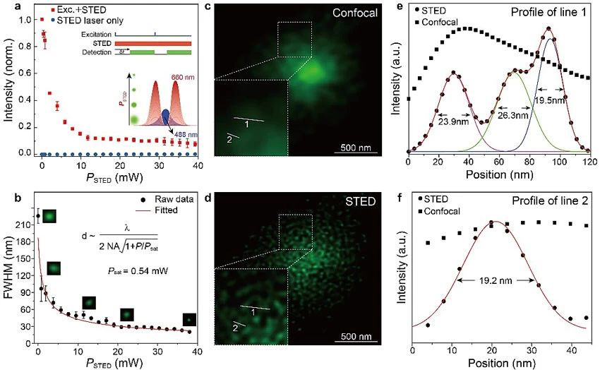
Recently, the team from Shenzhen University and the National University of Singapore (NUS) have collaborated to develop a novel STED fluorescent label based on organic-silica hybrid fluorescent nanodots. These new STED fluorescent nanodots have ultra-small physical sizes (<2 nm) and approximately 100% fluorescence quantum yield, demonstrating extremely outstanding super-resolution performance (as illustrated in the figure).

Link:https://onlinelibrary.wiley.com/doi/full/10.1002/anie.201912404
In recent years, the Biomedical Photonics Research Center at Shenzhen University has made significant progress in the development of new methods and techniques for super-resolution imaging using STED (Stimulated Emission Depletion) microscopy. These advancements aim to enhance the imaging performance of STED and explore new applications.
One notable advancement is the introduction of Coherent Optical Adaptive Techniques (COAT) into STED microscopy. This innovation greatly improves the imaging depth and spatial resolution of thick samples. The research findings were published in Photonics Research (2017, 5(3):176), a top-tier journal with an Impact Factor (IF) of 5.522 according to the Chinese Academy of Sciences Journal Citation Reports (JCR).
Another improvement involves the integration of genetic algorithms to enhance Adaptive Optics (AO) aberration correction. This results in faster and more accurate aberration correction for STED imaging in real-time. The research was published in Nanophotonics (2018, 7(12):1971), a top-tier journal with an IF of 6.908 according to the Chinese Academy of Sciences JCR.
Additionally, the team achieved remarkable results by utilizing perovskite quantum dots for STED imaging. This significantly enhances the photobleaching resistance of probes, enabling ultra-high resolution down to 20nm. The findings were published in Advanced Materials (2018, 30:1800167), a top-tier journal with an IF of 25.809 according to the Chinese Academy of Sciences JCR.
Furthermore, the team developed a novel type of carbon dots as STED probes, demonstrating outstanding super-resolution characteristics during STED imaging. These carbon dots achieve ultra-high resolution of 19.7nm in live cell imaging while maintaining excellent photostability and biocompatibility. The relevant results were published in Nano Research, a journal with an IF of 8.515 according to the Chinese Academy of Sciences JCR.
(Link:https://doi.org/10.1007/s12274-019-2554-x)
Moreover, the team employed techniques such as lifetime control and phase diagram analysis to develop methods for reducing excitation light power and improving spatial resolution. These findings were published in Journal of Biophotonics (2018, e201800315) and Nanoscale (2018, 10: 16252-16260), both reputable journals with IFs of 3.763 and 6.97 respectively according to the Chinese Academy of Sciences JCR, as well as in the upcoming publication of Laser & Photonics Reviews.
The research mentioned above received support from various funding sources including the 973 Program, National Key Research and Development Program, National Natural Science Foundation of China, as well as funding from Guangdong Province and Shenzhen City.


![]() Add : No. 3688, Nanhai Avenue, Nanshan District, Shenzhen, Guangdong Province
Add : No. 3688, Nanhai Avenue, Nanshan District, Shenzhen, Guangdong Province ![]() Email : cpoe@szu.edu.cn
Email : cpoe@szu.edu.cn ![]() Phone: 0755-26538735
Phone: 0755-26538735 ![]() Fax : 0755-26538735
Fax : 0755-26538735